Adult Stem Cell Therapy & Platelet Rich Plasma
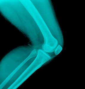
Anatomy of the Junction of the Vastus Lateralis Tendon and the Patella
 A contribution to Pub Med created by Hallisey MG, Doherty N, Bennett WF, and Fuklerson JP.
Forty-one knees from adult cadavera (twenty female and twenty-one male) were dissected to study the relationship between the longitudinal axis of the patella and the angles of insertion into it of the vastus lateralis and vastus lateralis obliquus muscles. The mean and variance in the angles of insertion of the vastus lateralis obliquus tendon were found to be significantly different between men and women (p less than 0.05 and p less than 0.01, respectively). Three distinct anatomical patterns in the insertion of the vastus lateralis obliquus muscle were delineated. The vastus lateralis muscle, particularly the vastus lateralis obliquus, creates an important lateral force-vector on the patella.
Click her for details of the report and to read the full text explaining this critical component of the anatomy of the knee.


